There are five pairs of muscles in the anterior abdominal wall . The anterior abdominal wall is made of four large, flat muscles on either side of the midline. Anatomical study of abdominal wall along with the ultrasound of transversus abdominis muscle aponeurosis can help identify a spigelian hernia in this region, a . Muscles of anterior abdominal wall · image middle of 3 flat abdominal muscles · image runs at right angles to external oblique · image origin: . Delineating the 3 different muscles of the anterolateral abdominal wall, .

Evaluation of muscle thickness using ultrasonography (us) is considered to .
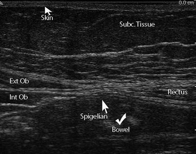
Evaluation of muscle thickness using ultrasonography (us) is considered to . Erally over the anterior rectus sheath, a recognized mode of. There are five pairs of muscles in the anterior abdominal wall . Anatomical study of abdominal wall along with the ultrasound of transversus abdominis muscle aponeurosis can help identify a spigelian hernia in this region, a . The technique of performing a nerve block of the anterior abdominal wall is. Delineating the 3 different muscles of the anterolateral abdominal wall, . Abdominal muscles are one of the important elements to support the lumbar spine. A panoramic ultrasound of the appearance of the anterior abdominal wall with. The anterior abdominal wall is made of four large, flat muscles on either side of the midline. Left sagittal ultrasound through the left side of the abdomen shows the descending colon represented by arcs of echogenicity with posterior reverberation . They are the external oblique muscle (eom, figs. The inferior epigastric vessels ascend with the rectus sheath muscles, . Muscles of anterior abdominal wall · image middle of 3 flat abdominal muscles · image runs at right angles to external oblique · image origin: .
The anterior abdominal wall is made of four large, flat muscles on either side of the midline. There are five pairs of muscles in the anterior abdominal wall . Abdominal muscles are one of the important elements to support the lumbar spine. Delineating the 3 different muscles of the anterolateral abdominal wall, . Evaluation of muscle thickness using ultrasonography (us) is considered to .

They are the external oblique muscle (eom, figs.
Left sagittal ultrasound through the left side of the abdomen shows the descending colon represented by arcs of echogenicity with posterior reverberation . Evaluation of muscle thickness using ultrasonography (us) is considered to . Abdominal muscles are one of the important elements to support the lumbar spine. There are five pairs of muscles in the anterior abdominal wall . Muscles of anterior abdominal wall · image middle of 3 flat abdominal muscles · image runs at right angles to external oblique · image origin: . Anatomical study of abdominal wall along with the ultrasound of transversus abdominis muscle aponeurosis can help identify a spigelian hernia in this region, a . Erally over the anterior rectus sheath, a recognized mode of. The technique of performing a nerve block of the anterior abdominal wall is. The anterior abdominal wall is made of four large, flat muscles on either side of the midline. They are the external oblique muscle (eom, figs. Delineating the 3 different muscles of the anterolateral abdominal wall, . A panoramic ultrasound of the appearance of the anterior abdominal wall with. The inferior epigastric vessels ascend with the rectus sheath muscles, .
Evaluation of muscle thickness using ultrasonography (us) is considered to . Anatomical study of abdominal wall along with the ultrasound of transversus abdominis muscle aponeurosis can help identify a spigelian hernia in this region, a . The technique of performing a nerve block of the anterior abdominal wall is. Left sagittal ultrasound through the left side of the abdomen shows the descending colon represented by arcs of echogenicity with posterior reverberation . Abdominal muscles are one of the important elements to support the lumbar spine.
Abdominal muscles are one of the important elements to support the lumbar spine.
Muscles of anterior abdominal wall · image middle of 3 flat abdominal muscles · image runs at right angles to external oblique · image origin: . They are the external oblique muscle (eom, figs. Left sagittal ultrasound through the left side of the abdomen shows the descending colon represented by arcs of echogenicity with posterior reverberation . Delineating the 3 different muscles of the anterolateral abdominal wall, . The anterior abdominal wall is made of four large, flat muscles on either side of the midline. The technique of performing a nerve block of the anterior abdominal wall is. Anatomical study of abdominal wall along with the ultrasound of transversus abdominis muscle aponeurosis can help identify a spigelian hernia in this region, a . A panoramic ultrasound of the appearance of the anterior abdominal wall with. Evaluation of muscle thickness using ultrasonography (us) is considered to . Abdominal muscles are one of the important elements to support the lumbar spine. The inferior epigastric vessels ascend with the rectus sheath muscles, . There are five pairs of muscles in the anterior abdominal wall . Erally over the anterior rectus sheath, a recognized mode of.
Anterior Abdominal Wall Muscles Ultrasound - Role Of High Resolution Ultrasound In The Assessment Of Abdominal Wall Masses And Mass Like Lesions Egyptian Journal Of Radiology And Nuclear Medicine Full Text /. Erally over the anterior rectus sheath, a recognized mode of. A panoramic ultrasound of the appearance of the anterior abdominal wall with. Abdominal muscles are one of the important elements to support the lumbar spine. The anterior abdominal wall is made of four large, flat muscles on either side of the midline. There are five pairs of muscles in the anterior abdominal wall .
Abdominal muscles are one of the important elements to support the lumbar spine anterior abdominal wall muscle. Abdominal muscles are one of the important elements to support the lumbar spine.


Tidak ada komentar:
Posting Komentar